What is AVN?

What is avascular necrosis?
Learn about how this complex hip condition.
What is AVN?
Hip avascular necrosis (AVN), also known as osteonecrosis of the hip aseptic necrosis or hip ischemic necrosis, is a condition in which the blood supply to the femoral head (the ball of the hip joint) is disrupted, leading to death of the bone tissue. Without proper blood flow, the bone tissue in the femoral head begins to die, leading to pain, stiffness, and limited mobility in the joint. Over time, the bone may collapse, resulting in further joint damage and the need for surgical intervention.
AVN may be spontaneous, or be caused by trauma, hemoglobinopathies, alcoholism, pancreatitis, or corticosteroid use. AVN only affects the ends of long bone, called the epiphysis. Although hip AVN is the most common form, you can also see AVN in different long bones such as in the metacarpal heads, metatarsal heads, humeral heads, the scaphoid or lunate (smalls bone in your wrist), or the talus (a small bone in the ankle).
Although AVN has been described as a relatively common condition, the incidence of hip avascular necrosis in Canada is not well established, however, it is estimated that approximately 10,000 to 20,000 Canadians are diagnosed with AVN each year, based on incidence rates in similar socioeconomic countries.1 Given the lack of comprehensive data on the incidence of hip AVN in Canada, it is important for healthcare professionals to remain vigilant in screening and diagnosing patients with symptoms of the condition. Early diagnosis and treatment can help prevent further joint damage and improve patient outcomes. Patients who suspect they may have hip AVN should consult with their healthcare provider to receive a proper diagnosis and develop a personalized treatment plan.
Imaging and Progression
The diagnosis of AVN is confirmed with imaging of the affected hip. In the earliest stages of the disease plain x-rays are often normal, however magnetic resonance image (MRI) studies can detect avascular necrosis earlier than x-rays.
The first radiographic findings include:
Increased joint space
Metaphyseal osteoporosis
Widened growth plate
Very small areas of AVN may improve without surgery, however progressive cases can lead to collapse of the head of the femur leading to deformity of the ball of the femur. This can affect the biomechanics of the hip, and present as a limp. Other radiographic signs such as crescent sign, rim sclerosis and patchy lucency can be observed with further progression of the condition. It may be necessary to verify the health of the opposite hip when evaluating avascular necrosis because studies have shown that the opposite hip may not hurt but may be affected up to 55% of the time within 2 years.2
The prognosis of AVN depends on:
Disease stage at the time of diagnosis (AVN caught and treated earlier have better outcomes)
Presence of other underlying conditions (such as alcoholism, blood disorders, history of AVN in other regions of the body, etc.)
Did you know that hip dislocations that are reduced within 12 hours have an AVN incidence of 22%; however, after 12 hours the incidence increases to 52%! Additionally, 15-50% of all femoral neck fractures develop AVN within 1 year.3
Factors associated with poor prognosis include:
Age over 50
Stage III+ at time of diagnosis
Greater than 1/3 of the bone’s weight-bearing area affected
Long history of cortisone treatment
Symptoms
Symptoms of avascular necrosis can be variable and often there is no pain at first. The most common symptom is vague non-specific hip pain, typically in the groin region when weight is placed on the hip (ex. standing up, getting from a seated to standing position, or getting out of bed in the morning). Other signs include muscle atrophy and decreased range of hip motion.
The pain can be felt in the groin area, the buttock area, and down the front of the thigh. As the problem progresses, the symptoms include development of a limp when walking and stiffness in the hip joint. Eventually, the pain can also be present at rest and may even interfere with sleep.
Stages
AVN has four stages that can progress over a period of several months to more than a year. The prognosis of AVN depends on the disease stage at the time of diagnosis and the presence of any underlying conditions. More than 50% of patients with AVN require surgical treatment within 3 years of diagnosis.4 Half of patients with subchondral collapse of the femoral head develop AVN in the opposite hip, so it is important that the other is assessed at time of the treatment.
The progression of AVN is categorized into stages based on the radiographic signs. Stage 1 looks normal on x-ray can be detected on MRI. Conversly, stage 2 can be detected on x-ray but there is no evidence of collapse of the femoral ball. Stage 3 signifies the collapse of the femoral head shown on x-ray. Stage 4 includes the collapse of the femoral head, but also signs of cartilage damage (osteoarthritis).
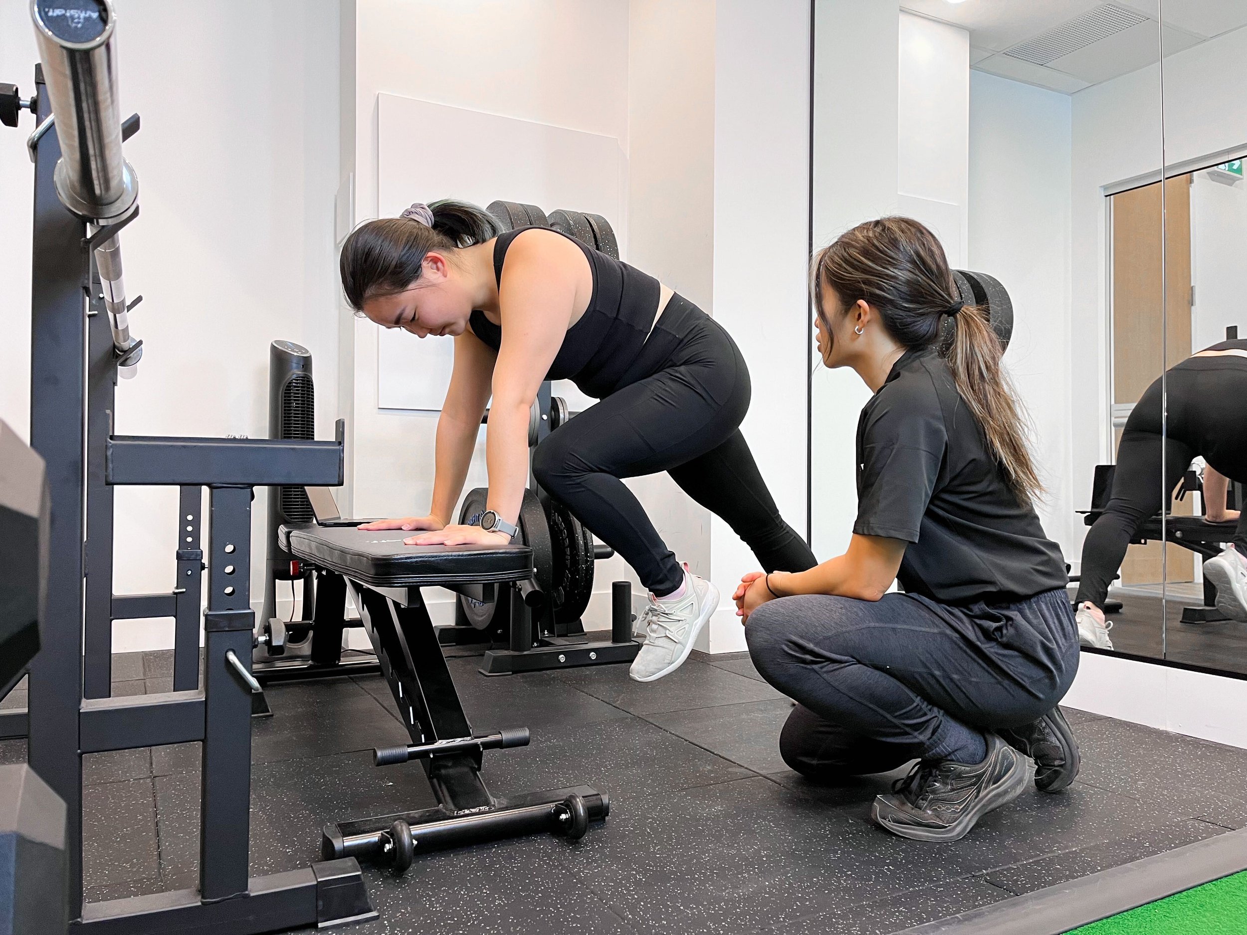
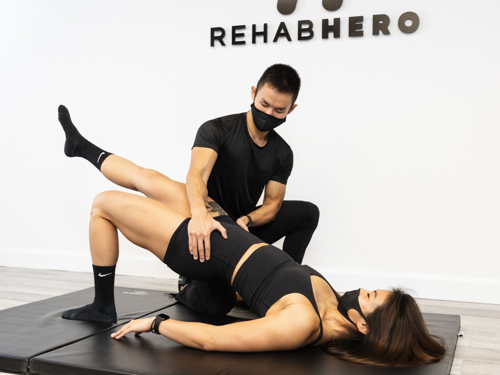
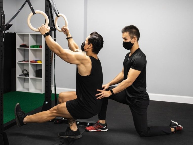
Conservative Therapy
Non-operative management may consist of protective weight-bearing (partial weight-bearing with crutches) for six weeks before re-evaluation. At this stage heat or ice can be used to manage pain. It is important to decrease the load put through the hip during weight-bearing as it gives the hip a chance to heal and modulate the pain.
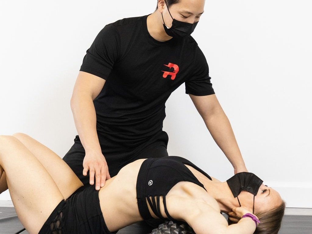
Soft tissue therapy of the gluteal region, low back, and anterior and lateral hip muscles may also provide relief. Use of an assistive walking device such as a cane or crutches can help with this. After full weight bearing is achieved, gait mechanics should be assessed to ensure that the other hip is not undergoing compensatory overload.
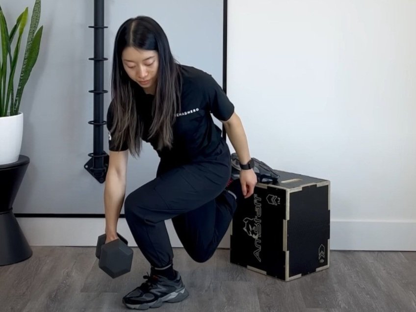
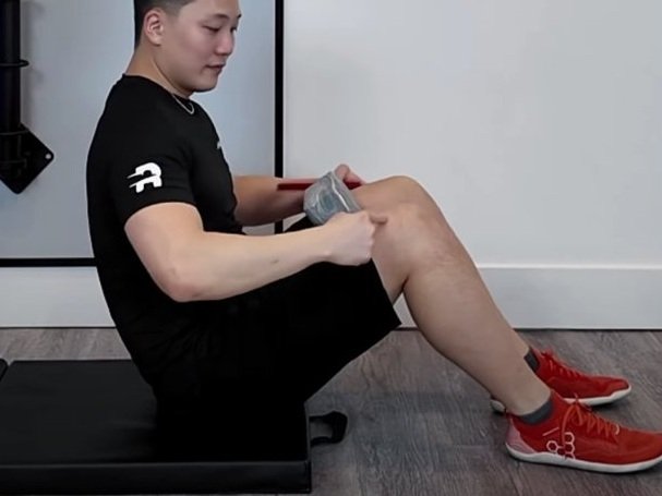
Within the clinic, mobilization of the hip, knees and low back should also be included to maintain their range of motion. Prescribed stretching exercises for at home management should be performed to maintain the range of motion and may also help in reducing swelling and improving blood flow to the healing areas. Compound exercises that involve the whole lower limb such as squats or lunge are functional examples of exercises as they assist with ADLs such as walking and climbing stairs. Balance and proprioceptive exercises are also important to avoid further decline in function, as a period of reduced mobility and weight bearing can negatively affect body position awareness.
If you think you or a loved one may have AVN, book in with your chiro or physio for a thorough examination. A history and physical examination can determine if further tests and imaging are required to confirm your diagnosis and rule out other pathologies. To book in with a chiropractor or physiotherapist at Rehab Hero click the book now button below.

Written by: Dr. Angeline Foote
Dr. Angie is a chiropractor in Markham that focuses on helping her patients through education, exercise, and hands-on manual therapy. Outside of the clinic you can find her rock climbing, working out, or participating in group classes.
References
“Diagnosis of Acute or Subacute Avascular Necrosis.” CADTH, www.cadth.ca/diagnosis-acute-or-subacute-avascular-necrosis. Accessed 8 May 2023.
Tripathy, Sujit Kumar et al. “Management of femoral head osteonecrosis: Current concepts.” Indian journal of orthopaedics vol. 49,1 (2015): 28-45. doi:10.4103/0019-5413.143911
Kellam P, Ostrum RF. Systematic Review and Meta-Analysis of Avascular Necrosis and Posttraumatic Arthritis After Traumatic Hip Dislocation. J Orthop Trauma. 2016 Jan;30(1):10-6. doi: 10.1097/BOT.0000000000000419. PMID: 26849386.
Sen, Ramesh Kumar. “Management of avascular necrosis of femoral head at pre-collapse stage.” Indian journal of orthopaedics vol. 43,1 (2009): 6-16. doi:10.4103/0019-5413.45318
Recent articles


Exercise videos