ITB Syndrome
How to heal ITB syndrome
Learn about this condition and how to start your recovery.
What is Iliotibial Band Syndrome
The Iliotibial band (ITB) is a thick cord of connective tissue that travels from the iliac crest to the lateral femoral condyle. The 2 main muscle groups that feed into the ITB include the tensor fascia latae (TFL) and gluteus maximus. Iliotibial band syndrome (ITBS) is typically an overuse injury resulting in pain and inflammation situated on the outside of the knee. Treatment interventions often include a combination of rest, load management, and active rehabilitation, with most cases fully resolving within 4-8 weeks.
Cause & Pathomechanics
Similar to other overuse injuries associated with endurance-based sports, ITBS is often a culmination of muscular imbalances, errors in load management, and faulty movement patterns. This can result in increased tension of the ITB, facilitating excess contact between its distal tendon and the lateral femoral condyle. Continued friction between the two surfaces leads to inflammation and pain which worsens with additional exertion. In the absence of sufficient recovery, inflammatory metabolites accumulate which result in increased local neovascularization and free nerve endings, which further propagates the pain cycle. Patients suffering from ITBS often report increased pain sensitivity around 30 degrees of knee flexion as there is maximal contact between the ITB and femur at this angle.
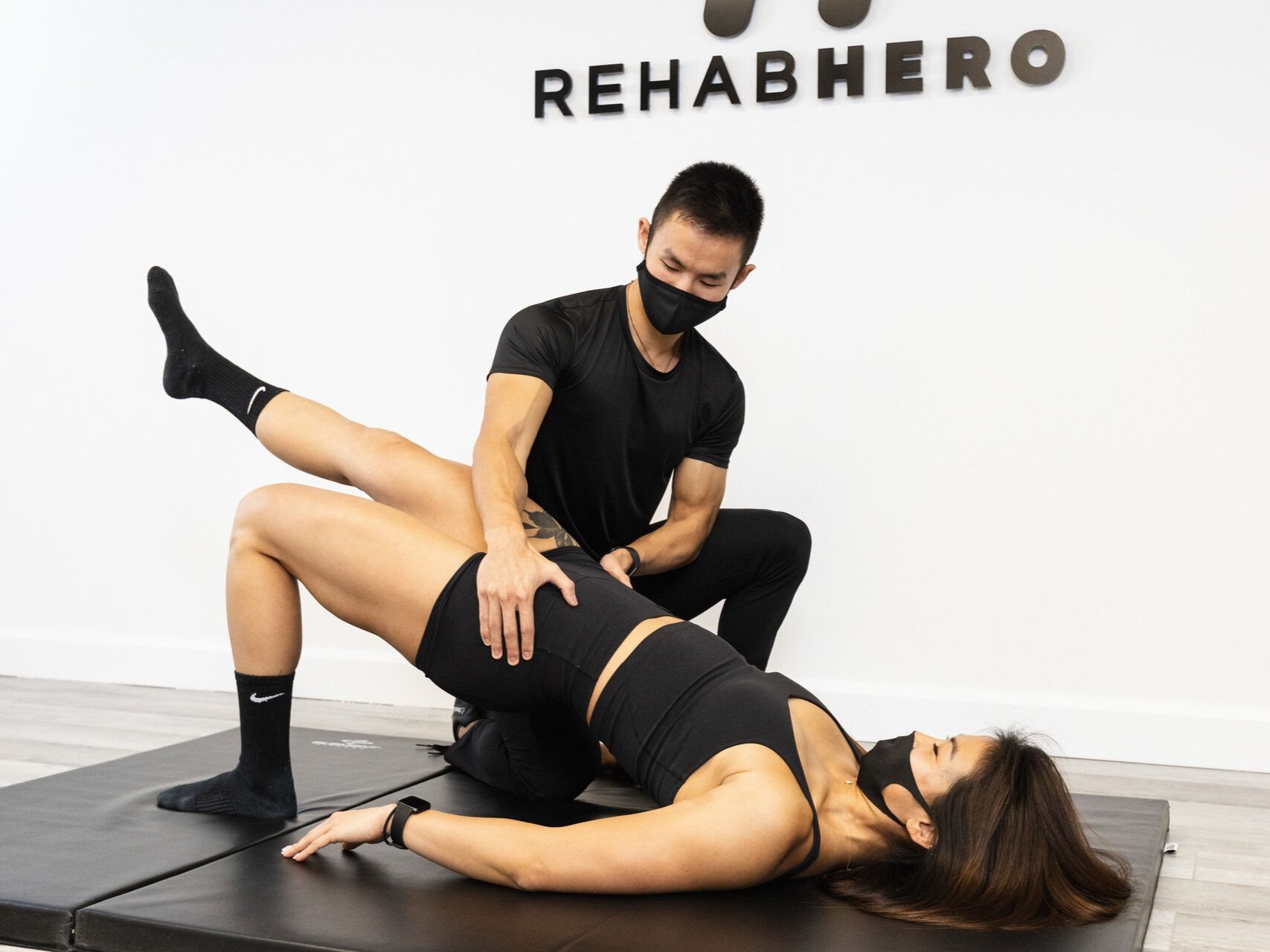
Weakness of the hip musculature, namely gluteus medius, may be present in cases of ITBS and contribute to the onset of symptoms. In ambulatory patterns such as walking and running, this muscle plays a pivotal role in maintaining pelvic stability and functional alignment of the lower extremities. However, if the gluteus medius is inhibited or deconditioned, the TFL may play a compensatory role in an attempt of re-stabilizing the pelvis. However, due to its attachment onto the ITB, continued overreliance on this muscle can ultimately result in excess tension and friction of the ITB over the lateral femoral condyle thus beginning the inflammatory cascade.
Changes in training variables may also contribute to pain and dysfunction. Athletes who rapidly increase their exercise volume may be especially susceptible to injury as the body does not yet have the prerequisite capacity of supporting the sharp increase in activity. Similarly, athletes who undergo changes in footwear or training surface without a sufficient acclimation period are also at risk of the training stimulus exceeding their body’s structural limits, thus leading to pain.
Furthermore, dysfunctional movement patterns also play an important role in symptom onset. Deficits such as excessive dynamic knee valgus, pelvic instability, and overpronation of the feet may lead to inefficient energy transfer through the kinetic chain. This ultimately results in elevated demands on the body and poorer shock absorption.
Common Symptoms
Pain and/or swelling situated at the outside of the knee associated with the onset of activity, worsening with continued exertion.
Pain that is exacerbated during uphill/downhill ambulation upon heel contact.
Pain that may be heightened at 20-30 degrees of knee flexion.
Snapping/crepitus-like sensation on the outside of the knee with walking, biking, and/or running.
Risk Factors:
A variety of risk factors may contribute to the onset of ITBS which include but is not limited to:
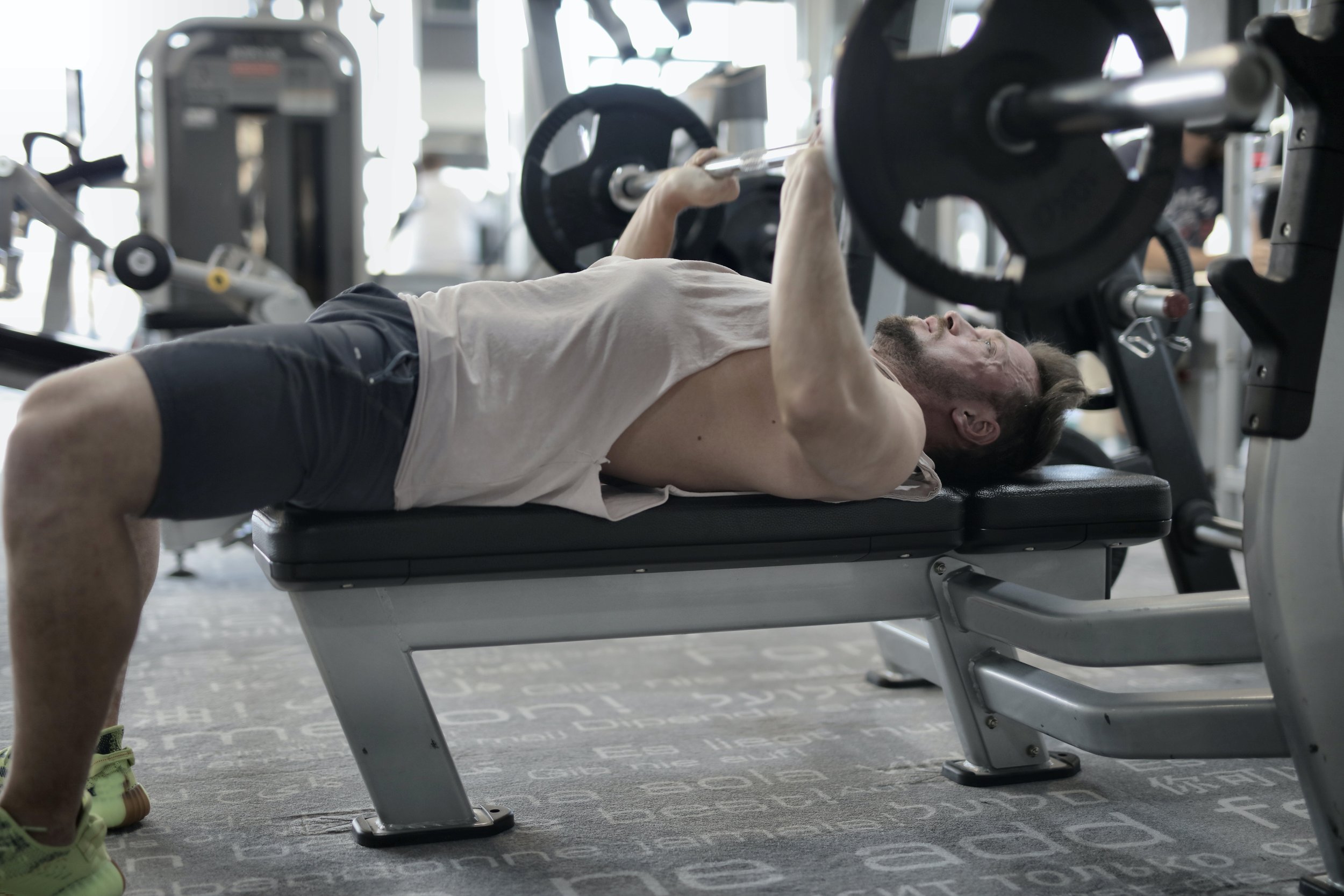
Endurance-based sports that require repetitive knee flexion-extension cycles such as running, cycling, basketball, soccer.
Anatomical variations that may include:
Genu varum/bow legs causing excess lateral tension of the ITB.
Leg length discrepancy resulting in uneven force absorption, pelvic asymmetry, and altered length tension relationships in the lower extremities.
Excess thickness/width of the ITB and/or a prominent lateral femoral condyle leading to increased risk of friction between the two tissues.
Muscular imbalances that may lead to the facilitation and/or inhibition of hip and core structures.
Improper footwear resulting in altered kinematics of the lower extremities and abnormal force absorption.
Adolescents undergoing rapid growth spurts causing asymmetrical growth between the muscular and skeletal systems.
How to treat ITB Syndrome
Rehabilitating ITBS will often begin with identifying relevant functional and logistical factors that contribute to excess tension in the ITB. In the short-term, implementing sufficient recovery periods, mobilizing tight tissues, and maintaining non-aggravating movement will help decrease inflammation and promote blood-flow. During this phase, other modalities may also be utilized to improve tissue healing. This may include:
1. Active release techniques to restore normal muscle tone/length of the hip musculature which decreases tension present in the ITB.
2. The graston technique and cupping can help decompress local tissues, improve perfusion, and remove adhesions.
3. Acupuncture to minimize pain, increase blood flow, and decrease inflammation.
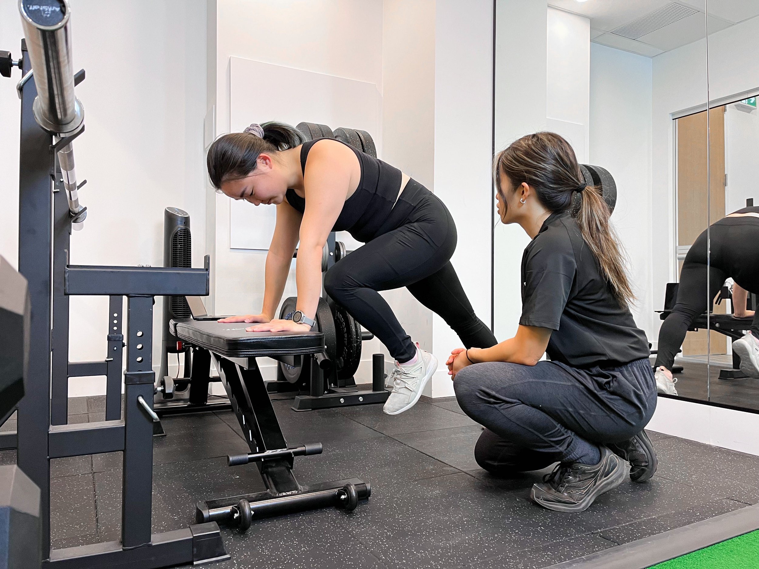
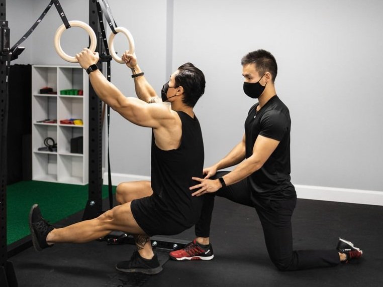
As acute symptoms subside, a progressive strength routine should be implemented with the goal of improving the capacity and activation of previously inhibited muscles. As tissue integrity increases, the need for compensatory patterns decreases, which reduces pain and dysfunction. Finally, retraining the nervous system to implement the newly activated musculature into the desired movement pattern is performed. This is done through activity-specific exercises and drills.
During the return-to-sport transition, a carefully titrated training program is critical. The individual will continue to progress their exercise volume and intensity in order to match the demands of their respective activity. At the same time, a journal may be utilized to track the individual’s pain pattern, objectively ensuring that overtraining is not occurring and sufficient recovery is taking place. Thus, the body is able to adapt to the increasing training demands while minimizing the risk of reinjury.
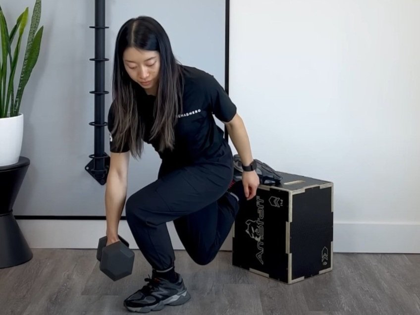
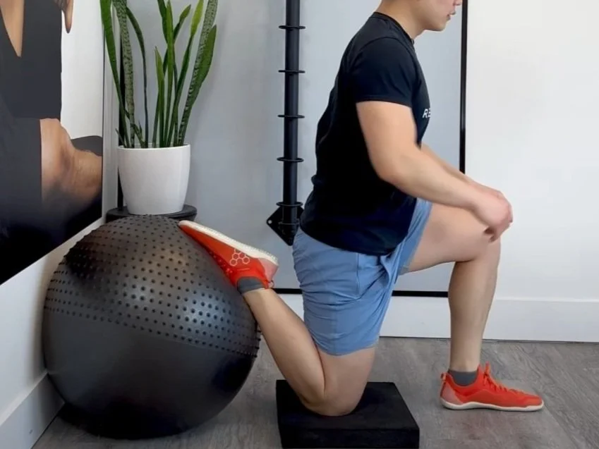
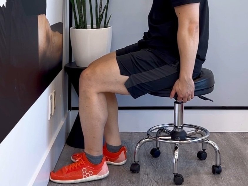
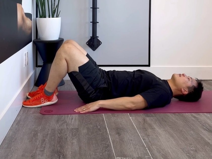
Beginner Exercises
You can watch a video made by Toronto chiropractor Dr. David Song below for a beginner program.

Here’s another video of his personal top 5 ITB syndrome exercises:

How Physiotherapy, Chiropractic, Massage Therapy, and Naturopathic Medicine Can Help
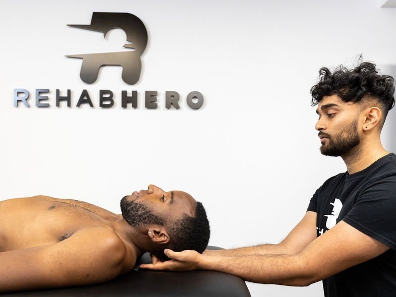
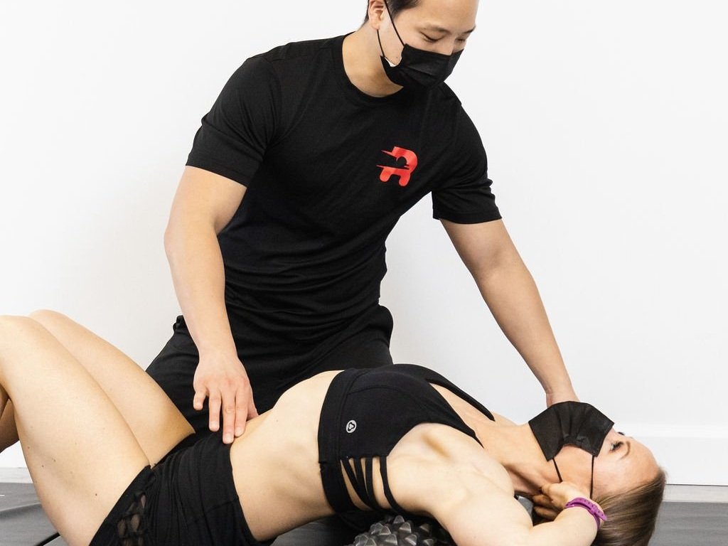
Having a licensed healthcare provider assess your knee can greatly accelerate your recovery and take the guesswork out of the process. With an in-depth postural and movement assessment, your clinician can accurately identify the source of your pain, relevant mobility limitations, and any compensatory movement patterns that contribute to the dysfunction. They will help create an individualized exercise program to enhance your tissue resilience and be there to guide you through every movement safely and effectively. A combination of manual therapy and mobilizations will be provided to restore motion to your muscles and joints. In addition, your clinician will also provide the necessary education to help monitor your recovery and prevent reinjury from occurring.
Contact us if you are suffering from knee pain or any other musculoskeletal issues. You can also click here to book in directly online with an experienced Rehab Hero chiropractor or physiotherapist.

Written by Eldon Thieu
Eldon is a physiotherapist that centers his practice in Markham. With a passion in optimizing weight lifting technique, he helps his patients excel in sports like CrossFit, Olympic weightlifting and bodybuilding injury free. Outside of the clinic you can find him playing beach volleyball, spike ball, or badminton.
Recent articles


Exercise videos