Wrist Pain & the Scapholunate Joint
Scapholunate Joint
Biomechanics, Anatomy, and Dynamic Instability Diagnosis
Anatomy & Pathomechanics of the Scapholunate Joint
The proximal carpal row, namely the scaphoid, lunate and triquetrum, have no tendons that insert unto them, and thus their motion is dependent on structures surrounding them via intrinsic, interosseous, and extrinsic carpal ligaments. Scapholunate injuries are the most common cause of carpal instability and can cause wrist dysfunction and interference with activities. Isolated injuries to the scapholunate interosseous ligament (SLIL) can cause abnormal joint mechanics, cartilage wear and tear, and degeneration. This ligament is c-shaped and attaches along the volar and articular surfaces dorsally and proximally, creating a crevice in the joint distally. This ligament can be subdivided into 3 regions comprised of different ligamentous material and anatomical properties. The dorsal region is the thickest, strongest, and most important stabilizer of the scapholunate articulation. It is considered a true ligament with transverse collagen fibers and restricts distraction, torsion, and translation. The palmar region is thinner, and restricts rotational movements. The proximal membranous region is consistent with fibrocartilage and does not contribute to restriction of motion.
The Ligaments of the Wrist & What They Do
The SLIL is considered to be the primary stabilizer of the scapholunate joint and possibly the entire carpal rows. It has great structural integrity, that when lost, compromises adjacent and surrounding ligamentous tissue. The surrounding ligaments provide secondary stability and have a little effect on scapholunate stability. However, compromised ligamentous stability in the surrounding structures can alter scapholunate kinematics. The radioscaphocapitate ligament, the long and short radiolunate ligament, and the radioscapholunate ligament have unknown interactions with the SL joint stability. The radioscapholunate ligament was previously regarded as a stabilizer, but has now been proven to work as a neurovascular conduit. The scaphotrapezial ligament complex is considered a secondary stabilizer of the scapholunate joint along with the dorsal radiotriquetral and dorsal intercarpal ligaments.
Anatomy and Biomechanics of the Scapholunate Interosseous Ligament & Rehab Theory
The SLIL is highly innervated by mechanoreceptors that provide fast input for the activation of antagonist muscles which include the extensor carpi radialis longus (ECRL), flexor carpi radialis, abductor pollicis longus (APL), and inhibits the extensor carpi ulnaris. It is these proprioceptive mechanisms that give rationale to proprioceptive training for rehabilitation. The role of the aforementioned muscles is important as isometric contractions can alter the position of the scaphoid. The ECRL and APL isometric contractions can create a supination moment on the scaphoid which may be protective of the dorsal SLIL, and better approximates the scaphoid to the lunate. Isometric contractions of the FCR can also create a supination moment on the scaphoid while pronating the triquetrum, creating another protective effect on the SLIL. Rehabilitation programs must be careful not to activate the Flexor Carpi Ulnaris and Extensor Carpi Ulnaris muscles as they produce a maximal cubital inclination of the scaphoid, which may increase the SL gap.
Coupled Movement Between the Scaphoid and Lunate
The scaphoid, lunate, and triquetrum rotate collectively in flexion and extension. In radial deviation, the scaphoid is driven into flexion by the distal carpal row and pulls the lunate passively with it through the strong SLIL. In ulnar deviation, the hamate causes the triquetrum into dorsal tilting via a screw-like mechanism, causing the scaphoid and lunate to rotate into extension through coupled rotation and ligament interactions into a dorsally translated position. Dorsal translation of the distal row causes tensioning of the radioscaphocapitate ligament which forces the scaphoid into extension. During wrist extension, the segments rotate as a unit, with extrinsic ligaments locking the scaphoid, lunate, and triquetrum to the capitate in extension.
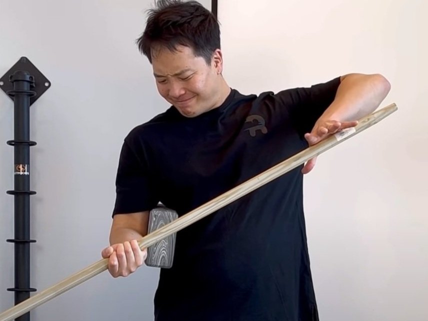
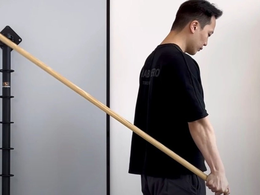
Scaphoid flexion exceeds lunate flexion by 35 degrees, and scaphoid pronation is 3 times that of lunate pronation during wrist flexion, and lunate ulnar deviation exceeds scaphoid deviation. Together, they form a coupled motion and functional arc commonly described as the Dart Throwing Motion (DTM), or radial deviation and extension, to ulnar deviation and flexion. This motion can be found in activities of daily living such as hammering, or pouring a glass of water. Since this is the functional movement pattern of the scapholunate joint, it is intuitive that rehabilitation of this joint recreates it.
What is Dynamic Scapholunate Instability?
Dynamic scapholunate instability is a concept that explains the symptoms of pain during mechanical loading or sudden shifts/ clunks experienced by patients without radiographic evidence of instability. Currently, dynamic scapholunate instability is considered a spectrum of injuries that can explain varying degrees of pain. It is currently defined by a wrist that is symptomatic during mechanical and load-bearing activities, demonstrating abnormal kinematics during motion.
Common Mechanisms of Injury
The most common mechanism of injury is a fall onto the hypothenar region of the hand causing disruption of the scapholunate ligaments. Studies have shown that mechanical failure of this ligament occurs with forcible wrist extension in ulnar deviation and supination. The patient will present with poorly localized tenderness, with pain preventing provocative testing of the surrounding ligamentous structures. Hemarthrosis is an observable sign that would suggest serious ligament injury. Sub-acute injuries are presented with a history of painful popping or clicking with activities, decreased grip strength, and well-localized tenderness on the dorsal surface of the scapholunate junction.
What are Some Tests for DSI?
Watson’s scaphoid shift test will be positive in even subtle cases of ligamentous instability. Normal scaphoid function is flexion and pronation during the test, but with ligamentous instability, the scaphoid will forced by digital pressure to move from the scaphoid fossa onto the dorsal articular lip of the radius, and often accompanied by pain. When digital pressure is relieved, the scaphoid will then shift back into the scaphoid fossa producing a clunk as it reduces. This test produces a false positive in one-third (1/3) of patients. Plain film radiographs may also be obtained to confirm the diagnosis of scapholunate instability. If plain film static radiographs are negative, yet clinical suspicion remains, stress radiographs may be obtained to confirm diagnosis.
Three Levels of Severity
Occult instability (pre-dynamic instability) is the mildest form of scapholunate instability and is often caused by a FOOSH injury. Often there is only a partial tear of the SLIL. There is a tendency for patients to not report this pain immediately, and will usually only report pain and dysfunction with mechanical loading, and may or may not progress to more serious forms of instability. The results of plain film radiograph (static or stress) and fluoroscopy may appear normal.
Subtotal or complete tear can occur with more forceful traumatic events, causing loss of integrity of the dorsal portion of the ligament. This tear may be accompanied with tearing of the extrinsic ligaments of the wrist. It is as this severity that the injury is referred to as dynamic instability, which will cause dysfunction within the joint and it’s kinematics. This injury may appear normal for weeks to months on static radiographs, and thus stress radiographs are necessary to confirm diagnosis.
Complete tear of the SLIL with a tear of 1 or more secondary ligaments will allow the scaphoid to rotate into flexion. This injury is now known as scaphoid dissociation, where the lunate rotates independently of the scaphoid. The lunate will remain in extension due to its articulations with the capitate and and triquetrum. This injury will be apparent on static radiographs and can become permanent with delayed surgical treatment due to changes in ligament lengths supporting the fallen scaphoid.
If left untreated, the injury may progress degeneratively into a condition known as Scapholunate Advanced Collapse (SLAC), which is further subdivided into four categories:
SLAC I is characterized by osteoarthritic (OA) changes of the scaphoid facet of the distal radius.
SLAC II has OA changes progressing towards the proximal radioscaphoid joint
SLAC III has OA changes invading the radial midcarpal joint
SLAC IV is involvement of the radiolunate joint and entire carpus, although the radiolunate joint is usually spared for years to decades due to its orientation with the lunate facet of the radius in all positions of rotation
Who can Test for SLIL?
You can directly consult your local sports physiotherapist or chiropractor for the assessment of your wrist. Your health care provider will collect a thorough history of your injury and combine it with a physical assessment to determine what is causing your wrist pain. Depending on initial assessment findings, they will refer you to see your medical doctor to acquire x-rays and/or stress x-rays of your wrist to look for instability of the scapholunate joint. Depending on the results of your x-ray, you may then be referred to your orthopaedic surgeon for a consult on whether or not surgery will be necessary (for severe cases). Otherwise, for milder cases, conservative treatment through modalities and rehabilitation exercises can start with your health care practitioner (physio or chiro).
Please remember there are many causes for wrist pain and an x-ray is often not necessary (or helpful) to diagnose them. This is why an examination by your clinician is necessary to first assess whether or not an x-ray will be helpful. Depending on assessment results, this may save you from unnecessary radiation exposure.
References
1. Kitay A, Wolfe S. Scapholunate instability: current concepts in diagnosis and management. The Journal Of Hand Surgery [serial on the Internet]. (2012, Oct), [cited November 7, 2017]; 37(10): 2175-2196. Available from: MEDLINE with Full Text.
2. Holmes M, Taylor S, Miller C, Brewster M. Early outcomes of ‘The Birmingham Wrist Instability Programme’: A pragmatic intervention for stage one scapholunate instability. Hand Therapy [serial on the Internet]. (2017, Sep), [cited November 7, 2017]; 22(3): 90-100. Available from: CINAHL Plus with Full Text.

Written By:
Dr. David Song, Chiropractor, Strength Coach
Recent Articles


Wrist Exercises