Patellar Dislocation
How to Recover from Patellar Dislocations
Background Information and First Steps To Function
What is a Patellar Dislocation?
Patellar dislocations occur when there is a loss of contact between the patella (a.k.a. the knee cap) and the patellar groove of the distal femur (groove in the thigh that the patella sits in). Dislocations usually occur laterally (outwards) and may be associated with chronic subluxation (joint dislocates and immediately relocates) or dislocation.
What is this condition known as?
This condition is also commonly referred to a patellofemoral pain syndrome (PFPS), dislocated kneecaps or patellar luxation.
Why do patellar dislocations occur?
There are two main reasons why the knee cap may dislocate. The most common cause of dislocation is due to indirect trauma. Indirect trauma occurs when the femur is internally rotated while the knee is in a knee valgus position (knocked knee). This typically occurs in sport while the foot is planted on the ground during pivoting motions.
The second and less common reason is due to direct trauma. Direct trauma occurs when the knee cap comes in direct contact with a lateral based blow that pushes it out of the patellar groove. This type of acute injury may lead to future chronic indirect dislocations.
Who typically experiences knee cap dislocations?
This type of dislocation is considered a common type of dislocation, typically occurring in females (2:1 ratio compared to males) aged between 12-25 years of age. Some other risk factors include:
Patellar Instability or Ligament Laxity
Previous history of patellar dislocation
Weak Vastus Medialis (1 of 4 quadriceps muscles)
Weak Vastus Medialis Oblique (1 of 4 quadricep muscles)
Tight Iliotibial Band (ITB)
Certain Sports (basketball, soccer - or anything that requires high velocity twisting of the knee)
Biomechanically patellar dislocations and subluxations are associated with:
Foot over pronation (pes planus - a.k.a. flat feet)
Knocked knees (Genu Valgum)
Femoral anteversion (internal rotation)
External tibial torsion
Elevated patella (patella alta)
Presentation of kneecap dislocation
This knee injury can sometimes mimic the symptoms of a knee sprain so be sure to contact a health care professional for an appropriate diagnosis. During the injury it is not uncommon to feel a grinding or tearing sensation and it is possible to hear a pop during the dislocation. A second pop may be heard during a subluxation (as the knee cap pops back into the patellar groove). It is also typical to be fear avoidant or apprehensive when it comes to moving the knee following an injury.
Who can diagnose a patellar dislocation?
You can get your knee pain diagnosed by any primary health care professional including your medical doctor, physiotherapist or chiropractor. If an acute dislocation is present, it is recommended to visit your medical doctor for relocation of the dislocated joint. Following relocation of the patella you may continue with conservative management with your physiotherapist or chiropractor.
Do I need an X-ray for diagnosis?
X-rays are not usually required to make a conclusive diagnosis of patellar dislocation, however it may be useful to rule out other conditions or to rule out any associated fractures. MRIs are not required either but may reveal soft tissue damage.
What are my treatment options?
For patellar dislocations you can see your massage therapist, chiropractor or physiotherapist for conservative treatment after the dislocation has been reduced.
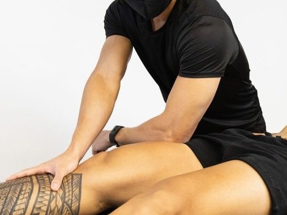
Massage therapy can be applied to compensating muscles to alleviate tension and pain. Typically it is applied to the hamstrings and quadriceps. Fascial techniques may also be used to allow for lymphatic drainage and to reduce edema. In chronic cases myofascial release therapy or deep tissue techniques may be applied to reduce muscle tension. This type of technique may be applied to the outer hip (gluteus minimus, gluteus medius, tensor fascia latae) and lateral quadriceps (vastus lateralis).
Acupuncture may be applied by your chiropractor or physiotherapist at local knee points to promote healing and to reduce pain.
Surgery is rarely required but may be necessary for chronic cases that have failed conservative therapy.
The main goal of treatment will be to increase knee stability and function for long term outcomes. This is best achieved when the aforementioned modalities are combined with a progressive rehabilitative exercise program.
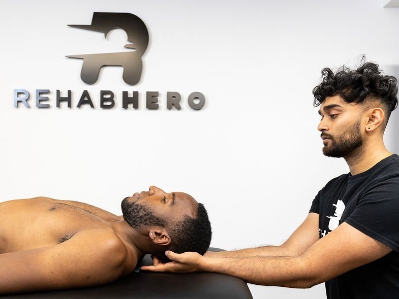
You can book in with a Rehab Hero therapist for an assessment of your knee using the button below:
Rehabilitation exercises for patellar dislocation
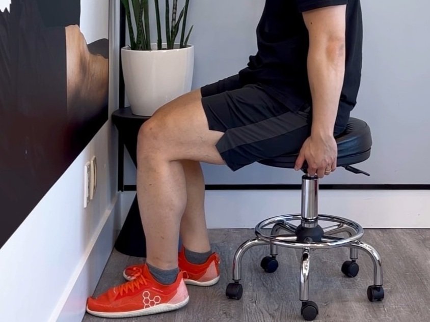
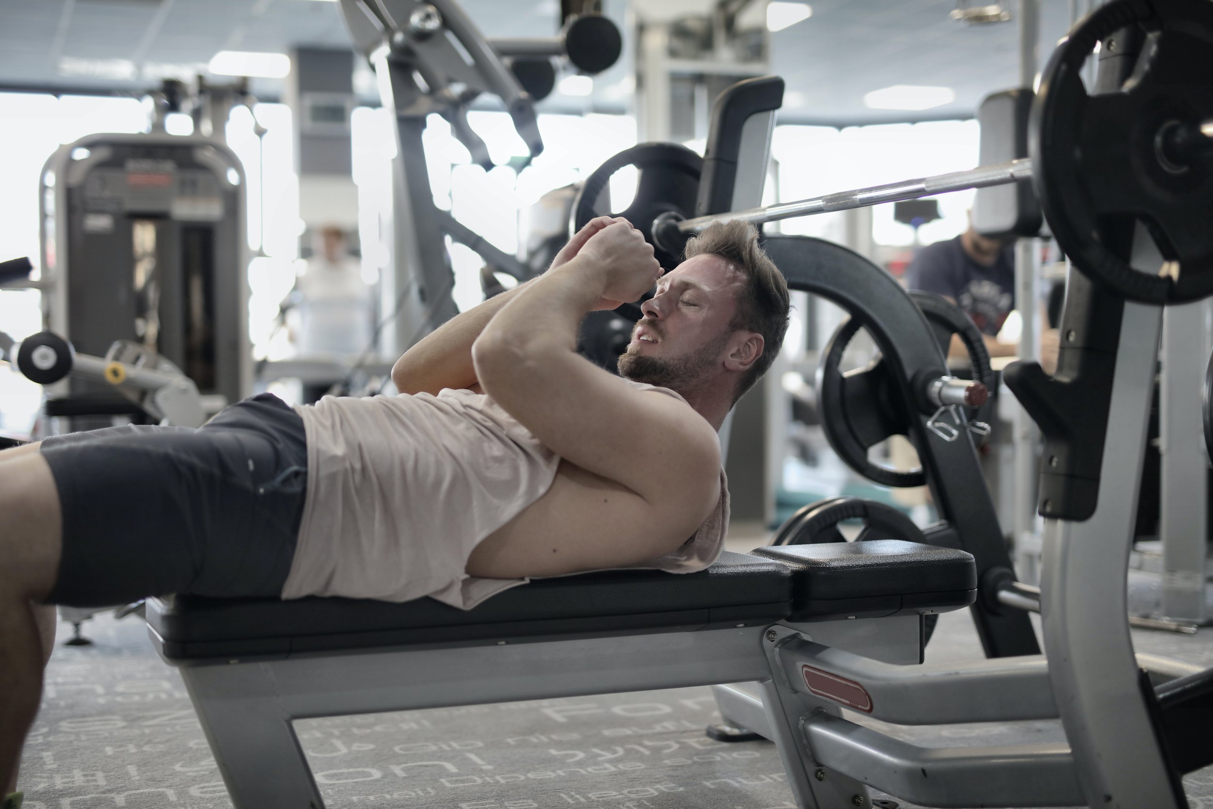
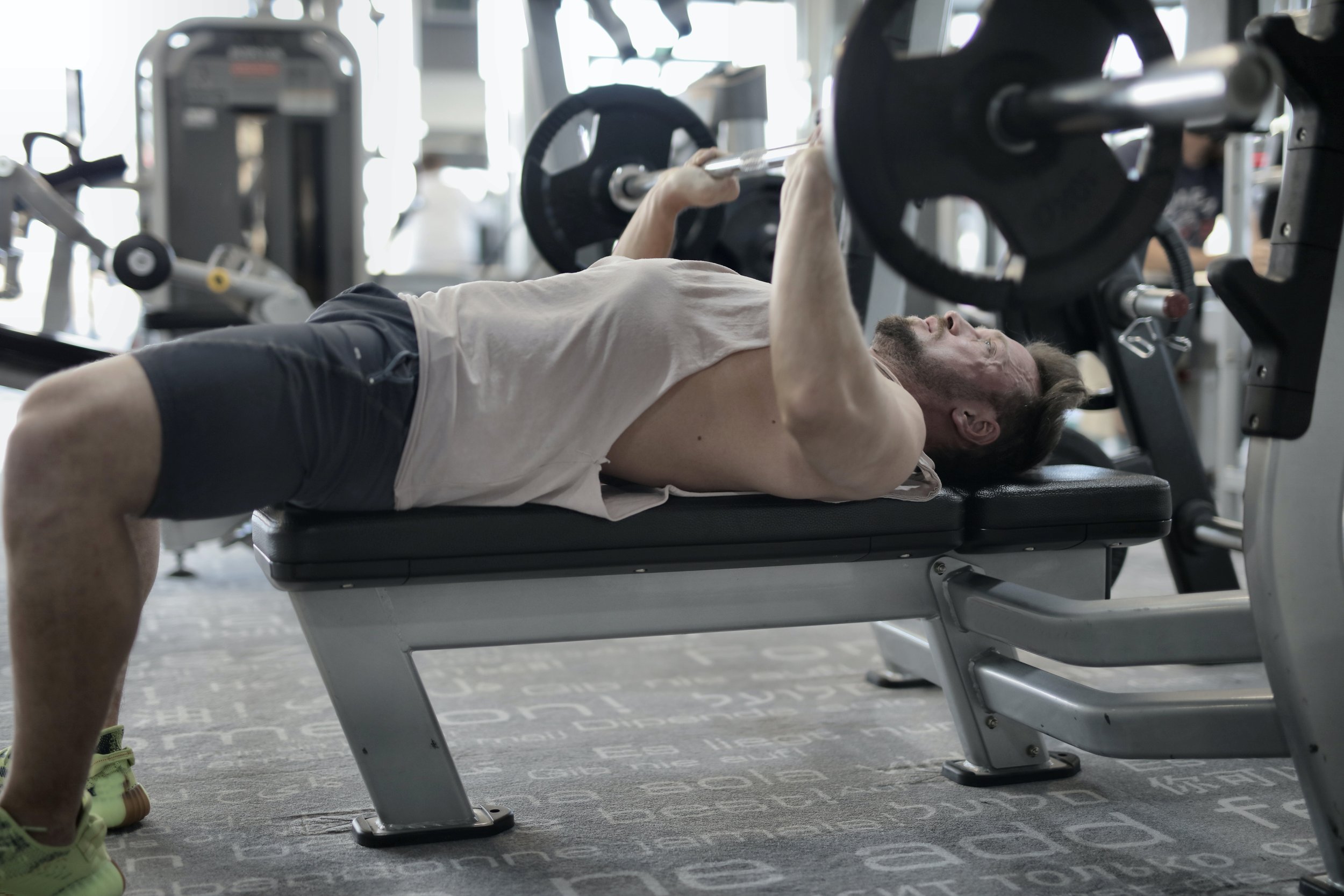
Passive or active range of motion exercise may start 4-7 days following the initial injury. The goal of these exercises is to reduce scar tissue formation and adhesion build up. It will also be important to complete range of motion exercises that also strengthen the quadriceps. An example includes the straight leg raise:

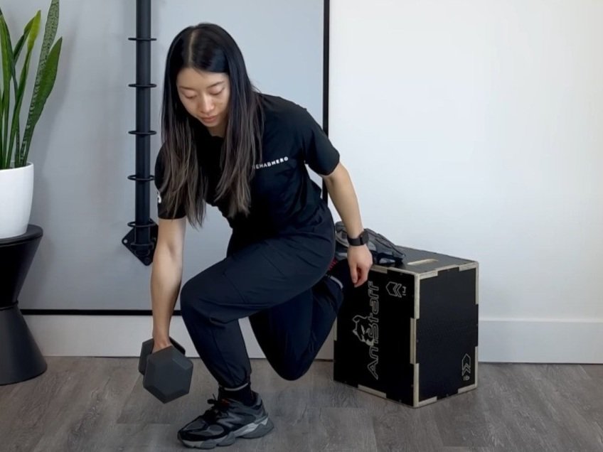
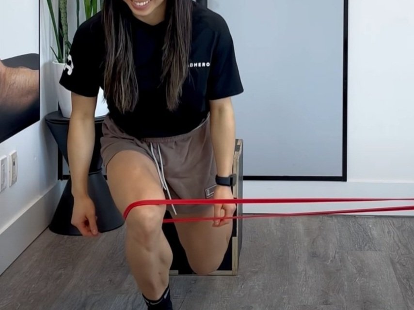
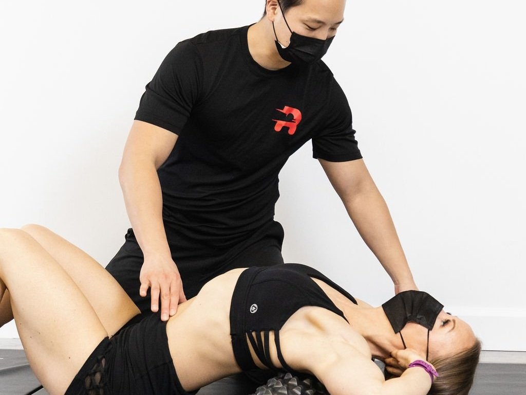
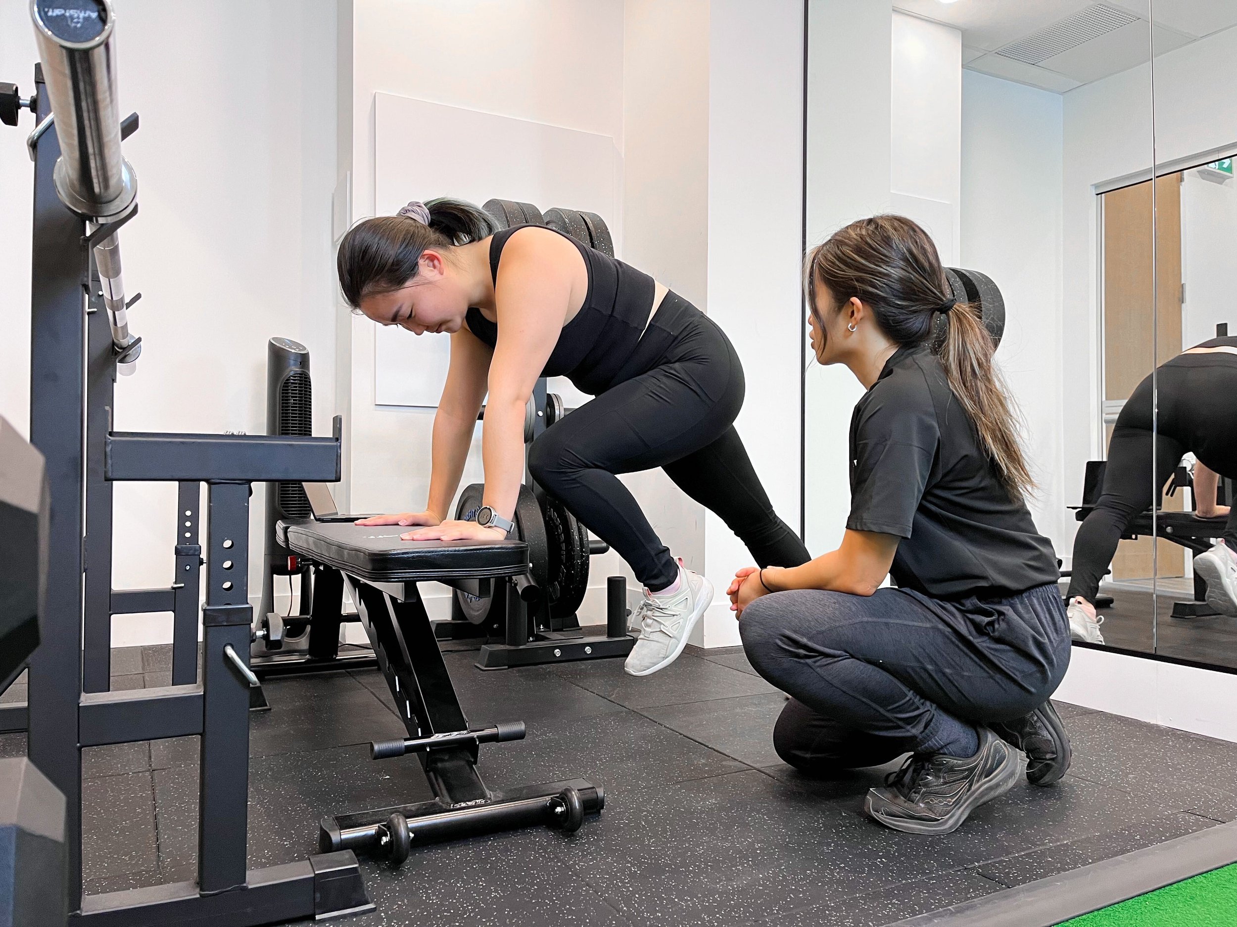
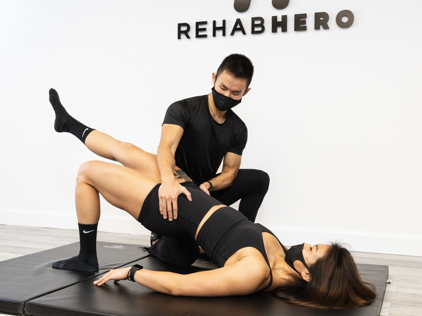
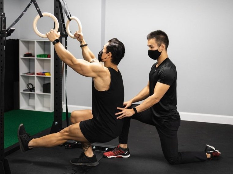
As sensitivity of the knee decreases following initial stages of rehabilitation (2 weeks after injury), progressive strengthening exercise and proprioceptive exercises may be completed to build patient confidence and reduce apprehension. An example of a strengthening exercise in a closed kinetic chain is the Step Up:

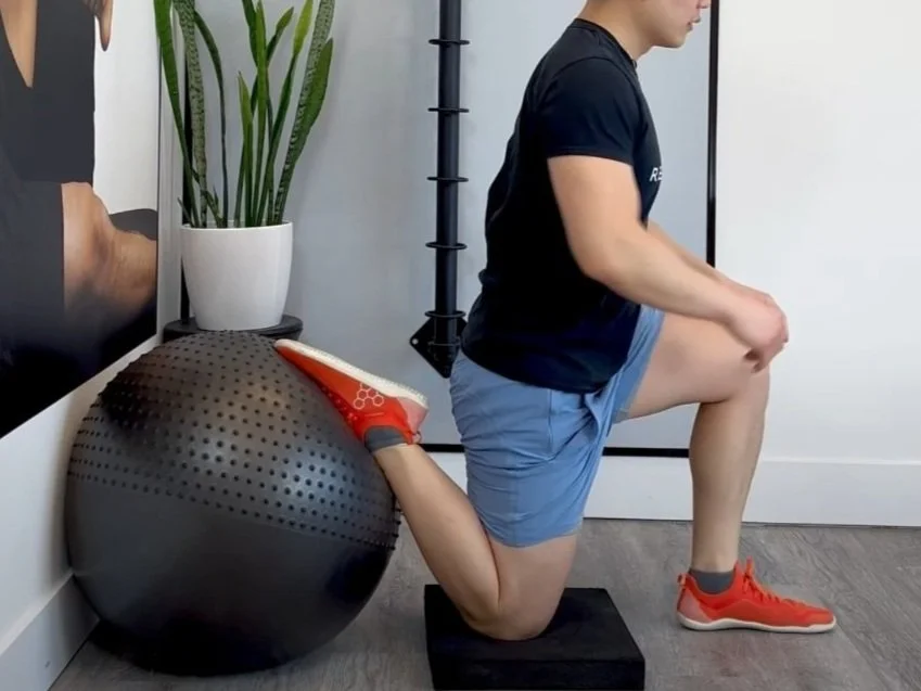
An example of a knee proprioception exercise is the Knee Orbit

What is the prognosis for this injury?
Overall prognosis is favourable with most responding well to conservative treatment. However there is a risk of a repeat dislocation during sport (roughly 50%). There is some research evidence suggesting that this condition may predispose degenerative joint disease later in life.

Written By:
Dr. David Song, Chiropractor, Strength Coach
Related Exercises


Quadriceps Exercises